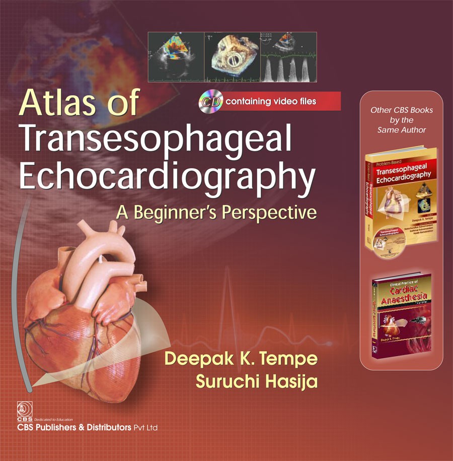
Transesophageal echordiography (TEE) has now become a standard monitoring tool inside a cardiac operating room. The present-day cardiac anesthesiologist is expected to be familiar with this imaging tool that has now become a part of the curriculum of the cardiac anesthesiology training program. Unlike most other imaging techniques, TEE is operator dependent and the quality of image produced (and its interpretation) depends on him. The first step in learning TEE is to be familiar with the normal and the common abnormal images. This atlas will go a long way in fulfilling this objective, its key features are:
Lucid illustration of normal as well as commonly encountered abnormal images.
Good quality images with brief and simple legends.
Brief description of echocardiography principles, valvular and congenital lesions along with other issues such as vegetations, masses, and thrombi.
A DVD containing video recordings of important normal and abnormal images has been provided.
The atlas is aimed at the beginners (anesthesiologists), however, trainee cardiologists and cardiac surgeons will also find it useful.

| Specifications |
Descriptions |
| ISBN |
9789386310651 |
| Binding |
NA |
| Subject |
Cardiology |
| Pages |
304 |
| Weight |
0.5 |
| Readership |
NA |


.png)
